Seeing Inside Your Arteries: Why It Matters
When a cardiologist performs an angioplasty, the goal is clear — open a blocked or narrowed coronary artery and restore healthy blood flow to the heart. Traditionally, this procedure has been guided by coronary angiography, which uses X-ray and a special dye to show the inside of the arteries as a two-dimensional silhouette.
Angiography is a powerful and proven tool. But it has a limitation: it shows only the outline of the artery's inner channel (the lumen), not the artery wall itself. Think of it like looking at the shadow of a pipe rather than the pipe from the inside. Sometimes, that shadow simply cannot tell the whole story.
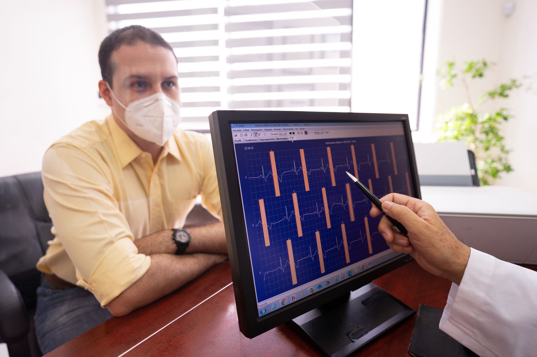
This is where IVUS (Intravascular Ultrasound) and OCT (Optical Coherence Tomography) come in. These two intracoronary imaging technologies give the interventional cardiologist a detailed, real-time "view from within" the artery — and they are changing the way angioplasty is performed for the better.
What Is IVUS?
Intravascular Ultrasound (IVUS) uses a miniature ultrasound probe mounted on a thin catheter. During the procedure, this catheter is gently advanced into the coronary artery. It sends out high-frequency sound waves that bounce off the artery walls and return as detailed cross-sectional images.
IVUS can show:
- The true size of the artery (which angiography often underestimates)
- The thickness and composition of plaque inside the artery wall
- Whether a plaque is stable or vulnerable (soft/lipid-rich vs. calcified)
- How well a stent has expanded and pressed against the artery wall
Because sound waves penetrate tissue deeply, IVUS is especially useful when the artery has heavy calcium deposits or when the cardiologist needs to understand how far plaque extends along the vessel.
What Is OCT?
Optical Coherence Tomography (OCT) works on a similar principle but uses near-infrared light instead of sound waves. Because light travels faster and has a much shorter wavelength than sound, OCT produces images that are 10–15 times sharper than IVUS.
With OCT, a cardiologist can clearly see:
- Thin layers of tissue as fine as 10–20 micrometres (far too small for angiography to detect)
- Thin-cap fibroatheroma — a particularly vulnerable type of plaque prone to rupture
- Tiny gaps or "malapposition" between the stent struts and the artery wall
- The precise edges of a stent — so there are no gaps left untreated
OCT's exceptional resolution makes it the preferred imaging tool when fine structural detail is needed, for example, when assessing why a previously placed stent is causing a problem or when placing a stent in a very complex lesion.
IVUS vs. OCT: A Quick Comparison
Both tools are valuable. They are not competitors — they are complementary. Here is a simple way to understand their differences:
| Feature | IVUS | OCT |
|---|---|---|
| Imaging method | Ultrasound (sound waves) | Near-infrared light |
| Image resolution | Moderate (100–150 µm) | Very high (10–20 µm) |
| Tissue penetration | Deep (useful for large vessels, calcium) | Shallower (best for fine surface detail) |
| Best use | Plaque sizing, vessel sizing, calcification | Stent assessment, thin plaque, edge detail |
| Contrast dye needed | No | A small flush is required |
In many complex procedures, an experienced interventional cardiologist may use insights from both modalities to plan and confirm the procedure.

How Do IVUS and OCT Make Angioplasty More Precise?
This is the heart of the matter (no pun intended). Here is how these imaging tools improve each stage of an angioplasty procedure:
1. Better Planning Before Stent Placement
Angiography may show a narrowing but not reveal why the narrowing exists — is it soft plaque, hard calcium, or a combination? IVUS can identify heavy calcification early, alerting the cardiologist that a special cutting balloon or rotational atherectomy device may be needed before placing a stent. This preparation helps the stent expand fully and sit properly.
2. Choosing the Right Stent Size
Undersized stents are a common cause of future problems — they can slip, underperform, or lead to re-narrowing. IVUS measures the true diameter and length of the artery far more accurately than angiography alone. For example, a 55-year-old patient with a vessel that appears 3 mm on angiography might actually measure 3.5 mm on IVUS — a difference that significantly affects stent selection.
3. Confirming Full Stent Expansion
After a stent is deployed, the cardiologist needs to confirm that it has opened fully and that every strut (the tiny metal segments of the stent) is in firm contact with the artery wall. OCT, with its high resolution, can show individual struts with remarkable clarity. Any areas of incomplete expansion or malapposition can be identified and corrected immediately — during the same procedure — rather than discovered weeks later.
4. Finding the Right Landing Zones
Every stent must start and end in healthy tissue. If a stent edge lands on a diseased segment of artery, that edge may become a new site of narrowing over time. IVUS and OCT help the cardiologist identify exactly where the healthy artery begins and ends, ensuring the stent covers the entire diseased area without unnecessarily extending into healthy tissue.
5. Investigating Stent Failure
If a patient returns with chest pain after a previous angioplasty, intracoronary imaging can reveal why the stent is not performing well — whether it is due to in-stent restenosis (tissue growing inside the stent), stent thrombosis (a clot), or stent fracture. This information directly guides the treatment decision.
What Does the Evidence Say?
Multiple large international studies have shown that using IVUS or OCT guidance during angioplasty is associated with:
- Better stent expansion, which reduces the risk of the artery re-narrowing
- Lower rates of major cardiac events (heart attack, need for repeat procedures) over the following years
- Improved long-term outcomes, particularly in complex lesions such as left main artery disease, long lesions, and bifurcation lesions
Guidelines from leading cardiology societies — including the European Society of Cardiology — now recommend considering intracoronary imaging guidance for complex angioplasty procedures.

Is This Available in Rajkot?
Advanced intracoronary imaging is no longer limited to large metropolitan hospitals. Interventional cardiologists in Rajkot and across Gujarat are increasingly incorporating IVUS and OCT into their catheterisation labs for complex cases. Patients undergoing angioplasty for complex or high-risk coronary disease can now benefit from this level of precision closer to home.
If you or a family member has been advised an angioplasty, it is perfectly reasonable to ask your cardiologist whether intracoronary imaging guidance is suitable for your specific case.
Key Takeaways
- IVUS uses ultrasound to provide cross-sectional images of the artery wall and plaque — excellent for sizing vessels and detecting calcium.
- OCT uses light waves to provide ultra-sharp images — ideal for assessing stent struts and fine plaque detail.
- Together, they help cardiologists plan better, place stents more accurately, and confirm results before closing the procedure.
- Both are non-invasive to the surrounding body (the catheter travels through the artery already being treated) and add minimal extra time to the procedure.
- Evidence supports that imaging-guided angioplasty is associated with improved long-term outcomes, especially for complex cases.
- These technologies are increasingly available to patients in Rajkot and Gujarat.
This article is intended for general patient education only and does not constitute individual medical advice. Every heart condition is unique. If you have questions about your coronary artery disease or an upcoming angioplasty, we warmly encourage you to book a consultation with a qualified interventional cardiologist — Dr. Nikhila Pachani and her team in Rajkot are here to guide you with clarity and care.
Frequently asked questions
- What is the difference between IVUS and OCT in angioplasty?
- IVUS (Intravascular Ultrasound) uses sound waves to image the artery wall and is excellent for measuring vessel size and detecting calcium. OCT (Optical Coherence Tomography) uses near-infrared light and produces much sharper images, making it ideal for assessing stent strut placement and fine plaque details. Both are used inside the coronary artery during the angioplasty procedure to guide stent placement.
- Is IVUS or OCT-guided angioplasty safe?
- Yes. Both IVUS and OCT are well-established, widely used technologies with strong safety records. The imaging catheter travels through the same artery being treated, requiring no additional puncture site. Your cardiologist will assess your individual case to determine whether intracoronary imaging is appropriate for you.
- Does imaging-guided angioplasty take longer than standard angioplasty?
- IVUS or OCT guidance adds a relatively small amount of time to the overall procedure. The benefit — greater confidence in stent sizing, placement, and expansion — is considered well worth this additional time, especially for complex coronary artery disease.
- Can I ask my cardiologist about IVUS or OCT before my angioplasty?
- Absolutely. It is always encouraged to ask your cardiologist about the tools and techniques planned for your procedure. If you have a complex coronary lesion, a long blockage, or have had a previous stent, intracoronary imaging guidance may be particularly relevant to discuss. Dr. Nikhila Pachani in Rajkot is happy to explain what is most suitable for your specific condition.