
What Is Restrictive Cardiomyopathy?
Your heart is a muscle that works like a pump. To pump blood forward, it first needs to relax and fill with blood. In most people, this happens smoothly with every heartbeat. But in a condition called restrictive cardiomyopathy (RCM), the heart muscle becomes abnormally stiff. It cannot relax and stretch properly, which means it cannot fill with enough blood — even though the pumping action itself may remain normal or close to normal.
Think of it this way: imagine trying to squeeze water into a rigid, half-open bottle instead of a soft, flexible pouch. The bottle may still squeeze water out fine, but getting enough in there in the first place is the real problem. That is essentially what happens in RCM.
Restrictive cardiomyopathy is one of the rarer forms of heart muscle disease. It is less common than dilated cardiomyopathy (where the heart enlarges and weakens) or hypertrophic cardiomyopathy (where the walls thicken). But it is important to understand because it can significantly affect quality of life and requires careful, specialised management.
What Causes the Heart Muscle to Become Stiff?
In many people with RCM, an abnormal substance or process causes the heart's inner walls to become rigid. Some of the known causes include:
- Amyloidosis — a condition where abnormal protein deposits build up in the heart tissue. This is one of the most common identifiable causes of RCM.
- Sarcoidosis — an inflammatory disease that can affect multiple organs, including the heart.
- Haemochromatosis — excess iron deposits in the body that can infiltrate the heart muscle.
- Certain inherited (genetic) conditions — some people inherit gene changes that make their heart muscle fibres stiffer over time.
- Radiation or certain medications — previous radiation therapy to the chest or some chemotherapy drugs can, over years, cause the heart muscle to stiffen.
- Idiopathic RCM — in a significant number of patients, no clear cause is ever found, and the condition is described as idiopathic (meaning unknown origin).
Understanding the underlying cause is crucial because, in some cases, treating the root cause — such as managing iron overload in haemochromatosis — can slow progression.

Recognising Restrictive Cardiomyopathy: Symptoms to Watch For
Understanding restrictive cardiomyopathy symptoms and management begins with recognising how the condition feels to a patient. Because the heart cannot fill adequately, blood can back up into the lungs and the rest of the body, causing a range of symptoms. These often develop gradually, and many people initially attribute them to ageing or general fatigue.
Common Symptoms
- Breathlessness (shortness of breath) — especially during activity, and later even at rest or when lying flat at night.
- Fatigue and low energy — because the heart is not delivering enough oxygen-rich blood to the body.
- Swelling in the legs, ankles, and feet — fluid backs up into the lower limbs.
- Swollen abdomen — fluid can also collect in the belly (ascites), causing bloating and discomfort.
- Reduced exercise tolerance — even a short walk or climbing a flight of stairs can feel exhausting.
- Heart palpitations or irregular heartbeat — the abnormal filling pressures can trigger rhythm disturbances.
- Dizziness or lightheadedness — due to reduced blood flow to the brain.
A Helpful Illustration
To illustrate: a 60-year-old person might visit their doctor in Rajkot saying they have felt unusually tired for six months and that their ankles are noticeably swollen by evening. They may not realise their heart is struggling to fill properly — they might assume it is just "old age." This is why awareness of these symptoms matters. Early evaluation can make a meaningful difference.
How Is Restrictive Cardiomyopathy Diagnosed?
Diagnosing RCM requires a combination of clinical assessment and investigations. A cardiologist will typically follow a stepwise approach:
1. Physical Examination
The doctor will listen to the heart and lungs, check for fluid in the legs and abdomen, and assess blood pressure and pulse patterns.
2. Echocardiogram (Heart Ultrasound)
This is often the single most informative test. An echocardiogram shows:
- The size and thickness of the heart chambers
- How well the heart relaxes and fills (diastolic function)
- The pumping function of the heart
In RCM, the heart chambers often appear normal in size or slightly smaller, but the pressures inside are elevated — a characteristic pattern that distinguishes it from other conditions.
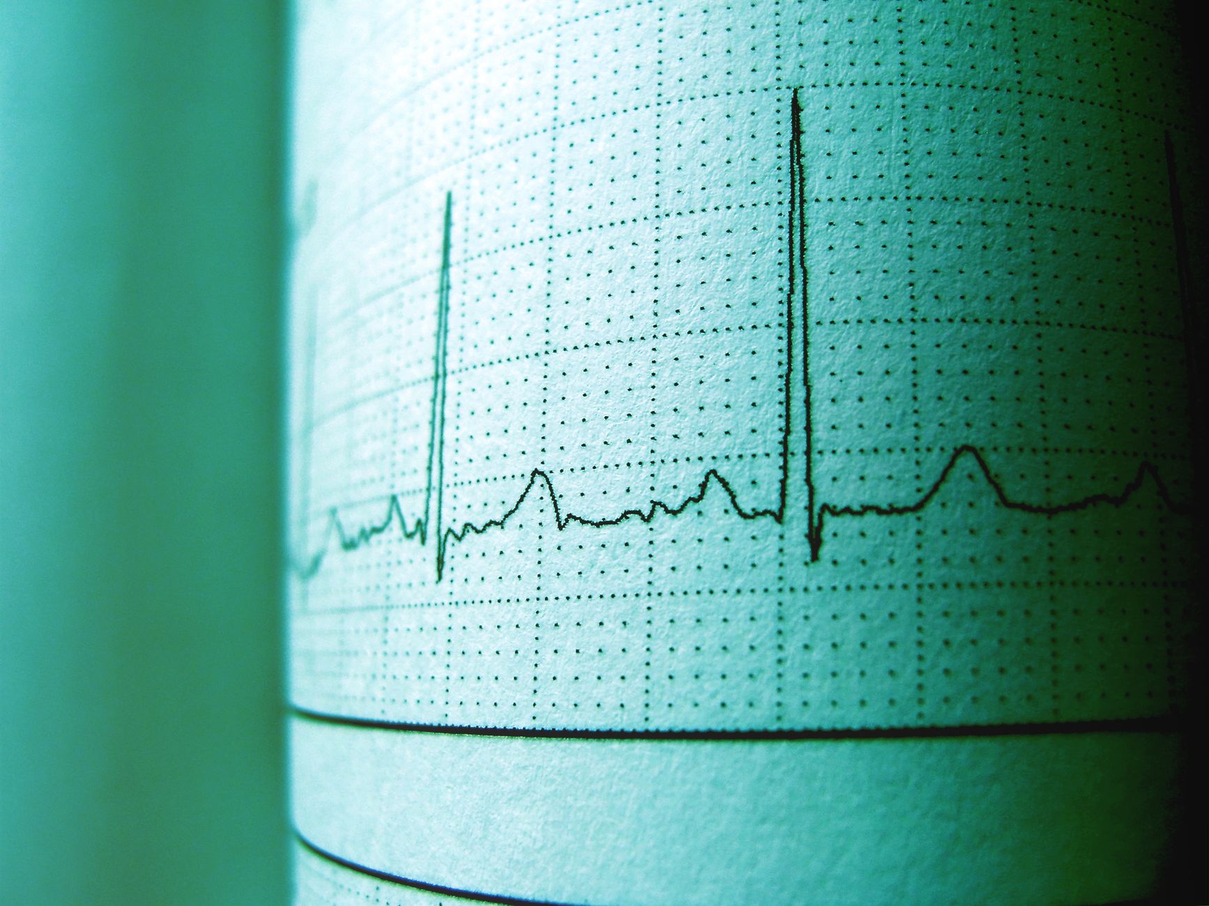
3. ECG (Electrocardiogram)
An ECG records the electrical activity of the heart. Certain patterns can suggest infiltrative diseases like amyloidosis — for example, low-voltage signals on ECG combined with a thickened-looking heart on echo is a red flag for cardiac amyloidosis.
4. Blood Tests
Tests including BNP or NT-proBNP (markers of heart stress), kidney function, liver enzymes, and specific tests for amyloidosis, iron levels, and inflammatory markers are ordered depending on clinical suspicion.
5. Cardiac MRI
Cardiac MRI provides detailed images of the heart muscle. It can detect areas of fibrosis (scarring) or infiltration that an echo may miss, and it is increasingly used in centres across India, including referral facilities available to patients in Rajkot.
6. Nuclear Imaging (Bone Scintigraphy)
This is a specialised scan particularly useful for detecting ATTR cardiac amyloidosis (a protein-related cause of RCM) without a tissue biopsy.
7. Biopsy
In selected cases, a small tissue sample from the heart (endomyocardial biopsy) may be needed to confirm the diagnosis and identify the exact type of infiltrating substance.
Management of Restrictive Cardiomyopathy
There is no single universal treatment for RCM because management depends significantly on the underlying cause. However, the goals are consistent: relieve symptoms, reduce fluid overload, protect the heart's rhythm, and — where possible — treat the root cause.
Managing Fluid Overload
Diuretics (water tablets) are often used to reduce the build-up of fluid in the lungs, legs, and abdomen. Careful dosing is essential — too little and the patient remains congested; too much can reduce the heart's already limited filling.
Treating the Underlying Cause
- Cardiac amyloidosis (ATTR type): Exciting advances have been made with medications like tafamidis, which stabilise the abnormal protein and have been shown to slow disease progression. This has changed the outlook for many patients.
- Haemochromatosis: Regular therapeutic phlebotomy (removing blood to reduce iron) or iron chelation therapy.
- Sarcoidosis: Anti-inflammatory treatments, including corticosteroids in some cases.
Managing Heart Rhythm Problems
Many patients with RCM develop atrial fibrillation (irregular heartbeat) or other rhythm disturbances. These are managed with rate-control medications, rhythm-control drugs, or blood thinners (anticoagulants) to reduce the risk of blood clots and stroke.
Lifestyle Adjustments
- Salt restriction — reducing dietary sodium helps prevent fluid retention.
- Fluid monitoring — some patients are advised to monitor and limit daily fluid intake.
- Regular, gentle activity — as tolerated, staying gently active supports overall wellbeing.
- Avoid alcohol — alcohol can worsen heart muscle function.
- Regular follow-up — frequent cardiology reviews are essential to adjust treatment as the condition evolves.
Advanced Options
In carefully selected patients — particularly younger individuals with idiopathic RCM — heart transplantation may be considered when other treatments are no longer sufficient. This decision involves a highly specialised evaluation at advanced cardiac centres.

Living With Restrictive Cardiomyopathy
A diagnosis of RCM can understandably feel overwhelming. But many people live meaningful, active lives with the right support. The key is:
- Early diagnosis — before significant complications develop.
- Understanding your specific cause — because targeted treatment is far more effective than generic care.
- Consistent follow-up with a cardiologist who knows your case well.
- Open communication about symptoms — never ignore new or worsening breathlessness, swelling, or palpitations.
Patients in Rajkot and across Gujarat have access to modern diagnostic tools — including echocardiography and advanced cardiac imaging — that allow accurate diagnosis and personalised management plans.
Key Takeaways
- Restrictive cardiomyopathy is a condition where the heart muscle becomes stiff, preventing the heart from filling properly with blood.
- Common symptoms include breathlessness, fatigue, leg swelling, and reduced exercise capacity.
- Diagnosis involves echocardiogram, ECG, blood tests, cardiac MRI, and sometimes biopsy.
- Management depends on the underlying cause — treating conditions like amyloidosis or haemochromatosis can meaningfully improve outcomes.
- Diuretics, rhythm management, lifestyle changes, and regular follow-up form the backbone of day-to-day care.
- New treatments (such as tafamidis for ATTR amyloidosis) have improved the outlook for certain types of RCM.
- Early evaluation and specialist care make a significant difference — do not dismiss persistent breathlessness or swelling as just ageing.
If you or a loved one are experiencing unexplained breathlessness, fatigue, or swelling, we warmly encourage you to seek a professional cardiac evaluation. You are welcome to book a consultation with Dr. Nikhila Pachani, Consultant Interventional Cardiologist, Rajkot — early assessment is always a wise step for your heart health.
Frequently asked questions
- What is the difference between restrictive cardiomyopathy and other types of cardiomyopathy?
- In restrictive cardiomyopathy, the heart muscle becomes stiff and cannot relax and fill properly with blood, even though the pumping function may remain relatively normal. In dilated cardiomyopathy, the heart enlarges and the pumping becomes weak. In hypertrophic cardiomyopathy, the heart walls become abnormally thick. Each type has different causes, symptoms, and treatment approaches.
- Can restrictive cardiomyopathy be treated or reversed?
- Whether it can be treated or reversed depends on the underlying cause. For example, managing iron overload in haemochromatosis or using newer medications for ATTR cardiac amyloidosis can slow or partially address the condition. In idiopathic cases (unknown cause), treatment focuses on relieving symptoms and preventing complications rather than reversing the stiffness. A specialist cardiologist can guide the most appropriate plan for each individual.
- What symptoms of restrictive cardiomyopathy should prompt an urgent cardiology visit?
- You should seek prompt medical attention if you experience sudden or rapidly worsening breathlessness, severe leg or abdominal swelling, new palpitations or irregular heartbeat, fainting or near-fainting episodes, or chest discomfort. If any existing symptoms worsen noticeably, it is always better to have them assessed sooner rather than later.
- How is restrictive cardiomyopathy diagnosed without a biopsy?
- In many cases, a combination of echocardiogram, ECG, cardiac MRI, blood tests, and specialised nuclear scans (such as bone scintigraphy for amyloidosis) can provide a confident diagnosis without the need for a heart biopsy. A biopsy is reserved for situations where non-invasive tests do not give a clear enough answer to guide treatment.