When Advanced Imaging Changes Everything
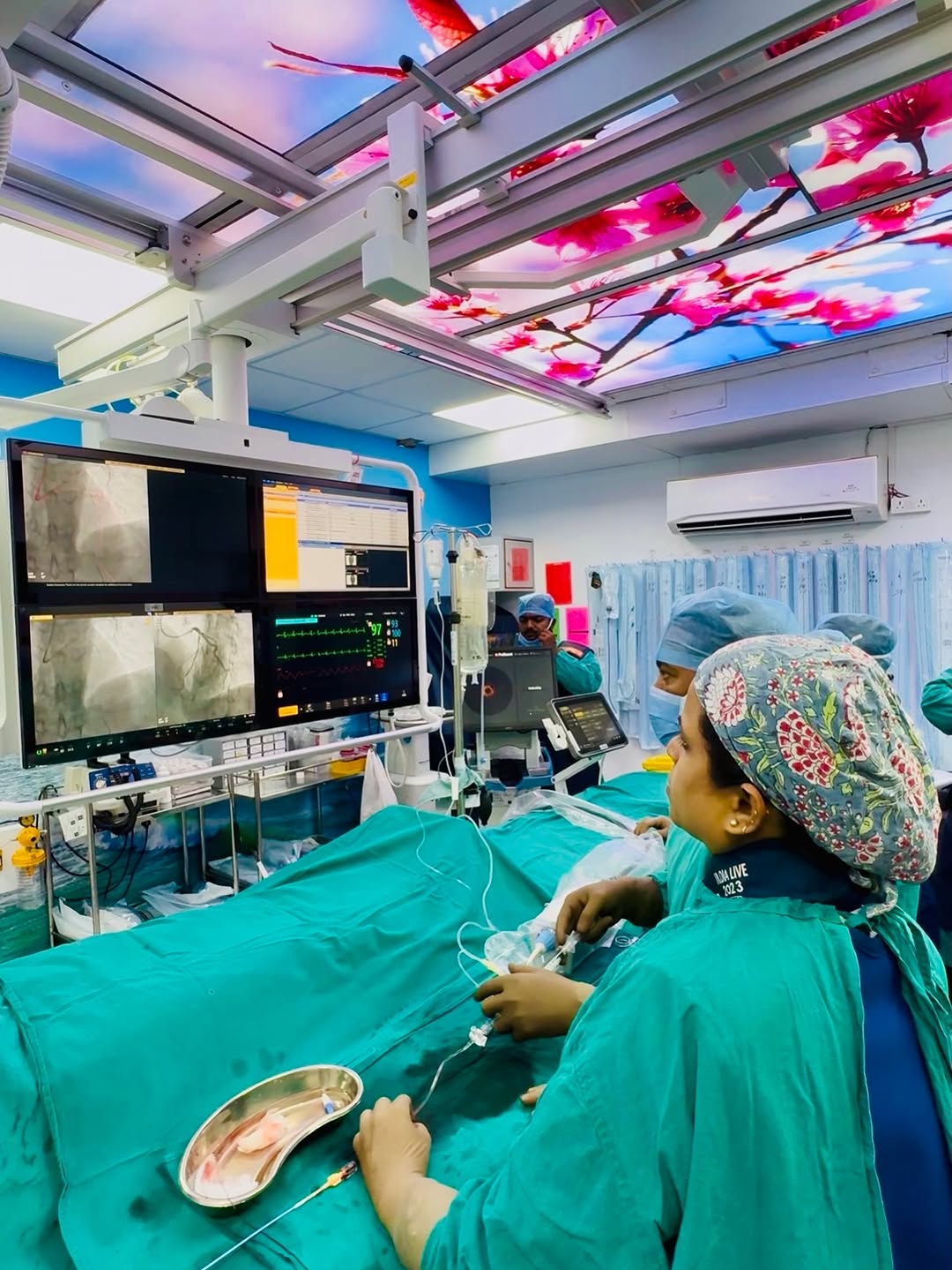
In a recent case at Backbone Medicity Hospital in Rajkot, Dr. Nikhila Pachani — Consultant Interventional Cardiologist — used a powerful imaging tool called Optical Coherence Tomography (OCT) to make a key decision about a patient's heart treatment.
What the Angiography Showed
The patient's coronary angiography revealed what appeared to be a critical lesion at the ostium (opening) of the Left Anterior Descending (LAD) artery — one of the most important arteries of the heart. Based on this finding alone, the initial plan was to go ahead with a Percutaneous Coronary Intervention (PCI), commonly known as angioplasty or stenting.
What OCT Revealed
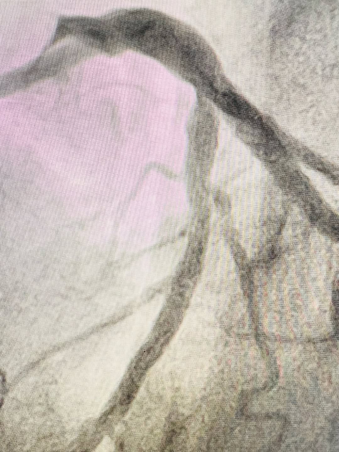
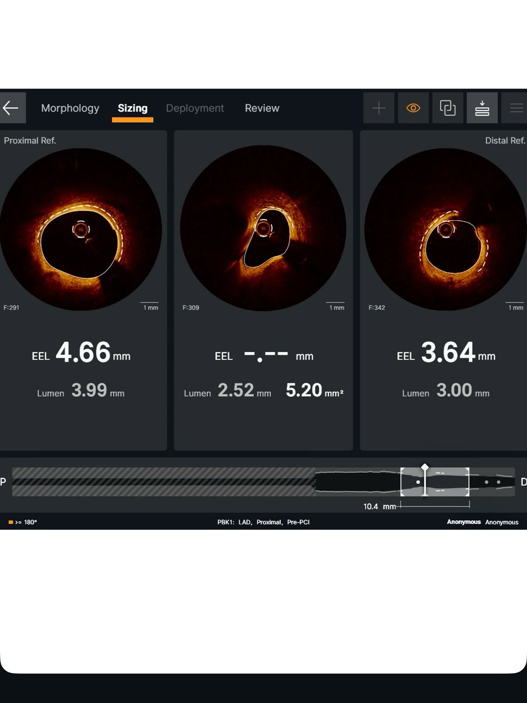
Before proceeding, Dr. Pachani performed an OCT scan — a high-resolution intracoronary imaging technique that looks inside the artery wall with great detail. The OCT images told a different story:
- The lumen (the space inside the artery) was adequate in size.
- There were no high-risk features such as thin-cap plaques or significant blockage.
- The lesion was ultimately classified as not significant enough to require intervention.
As a result, the treatment plan was completely changed — from planned stenting to optimal medical management (medicines and lifestyle changes).
Why This Matters for Patients
Angiography is a very useful test, but it only shows the outline or silhouette of an artery. It cannot always tell the doctor whether a narrowing is truly dangerous. OCT and similar intracoronary imaging tools look inside the artery wall, giving a much clearer picture of what is actually happening.
Using this technology helps doctors:
- Avoid unnecessary procedures that carry their own risks
- Personalise treatment based on accurate information
- Give patients the safest and most suitable care
"Angiography shows the outline, but intracoronary imaging reveals the truth." — Dr. Nikhila Pachani
A Step Towards Smarter, Safer Heart Care in Gujarat
This case is a good example of how advanced cardiac imaging is changing the way heart conditions are managed — not just in large metro cities, but right here in Rajkot and across Gujarat.
If you or a family member has been advised an angiography or heart procedure, speak with a qualified interventional cardiologist to understand all your options, including whether advanced imaging may help guide your treatment.