If your doctor has recommended a 2D echo test, you may have questions. What exactly is it? Will it be uncomfortable? What will it tell the doctor about your heart? You are not alone — many patients in Rajkot and across Gujarat ask these same questions every day. This article answers them in simple, clear language so you feel informed and confident before your appointment.
What Is a 2D Echo Test?
A 2D echo test — short for two-dimensional echocardiogram — is a type of ultrasound scan of the heart. Just like an ultrasound is used to look at a baby during pregnancy, a 2D echo uses sound waves to create moving pictures of your heart in real time.
The word "echo" comes from echocardiography, which simply means using sound echoes to study the heart (cardia = heart, graphy = recording). Unlike an X-ray or CT scan, a 2D echo does not use radiation. It is a non-invasive test — nothing enters the body, and most patients find it quite comfortable.
The "2D" refers to the two-dimensional images the test produces. These images allow the cardiologist to see the heart's structure and movement as it actually beats, giving far more information than a still picture ever could.
How Is the Test Performed?
The procedure is straightforward and typically takes between 20 and 40 minutes.
- You lie down on an examination table, usually on your left side, which brings the heart slightly closer to the chest wall.
- Small electrode stickers (ECG leads) are placed on your chest to record your heart rhythm at the same time.
- A gel is applied to the chest. This gel helps the sound waves travel smoothly between the probe and your skin — it may feel cool, but it is completely harmless.
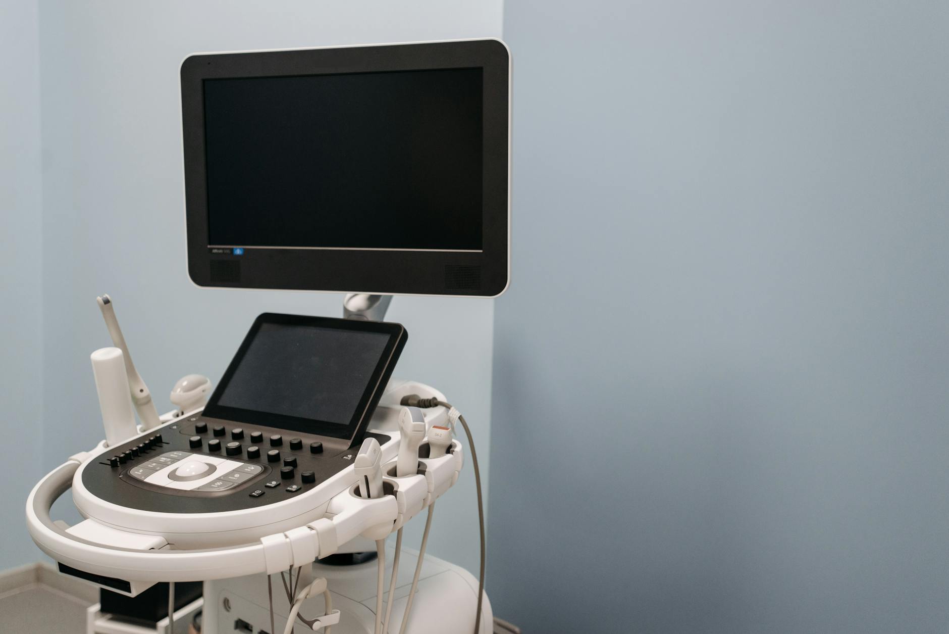
- A probe (transducer) is gently moved across different areas of your chest. The probe sends sound waves into the chest and picks up the echoes that bounce back from heart tissue.
- Images appear on a screen in real time. The cardiologist or sonographer watches and records these images for later analysis.
No needles, no dye, and no special preparation is usually needed — though your doctor may ask you to avoid eating for a few hours if a specific type of echo is planned.
What Does a 2D Echo Test Show?
This is where the 2D echo test truly shines. In a single session, it can reveal a remarkable amount of detail about your heart's health. Here is what your cardiologist looks at:
1. Heart Size and Shape
An enlarged heart (cardiomegaly) or an unusually small or thickened chamber can be spotted immediately. Conditions like hypertrophic cardiomyopathy (thickening of the heart muscle) are often first identified on a 2D echo.
2. Heart Pumping Function (Ejection Fraction)
One of the most important numbers your 2D echo provides is the ejection fraction (EF) — the percentage of blood pumped out of the heart's main chamber (left ventricle) with each beat. A normal EF is generally between 55% and 70%. A lower value may indicate heart failure or weakened heart muscle, while a very high value can signal its own set of concerns.
3. Heart Valves
Your heart has four valves that open and close with every heartbeat to keep blood flowing in the right direction. A 2D echo — especially when combined with Doppler technology — shows whether valves are:
- Narrowed (stenosis): Not opening wide enough, making the heart work harder.
- Leaky (regurgitation): Not closing properly, allowing blood to flow backward.
Valve problems are common in Gujarat's population, including rheumatic heart disease — a condition that can damage valves years after a strep throat infection in childhood.
4. Heart Wall Movement
The cardiologist examines every segment of the heart wall to check if it is moving normally. Areas that move poorly or not at all may indicate a previous heart attack or current lack of blood supply (ischaemia).
5. Fluid Around the Heart (Pericardial Effusion)
A healthy heart is surrounded by a small amount of fluid in the pericardial sac. If too much fluid accumulates — a condition called pericardial effusion — it can press on the heart. A 2D echo can detect even small amounts of this fluid quickly and reliably.
6. Blood Clots or Masses
Clots inside the heart chambers, or unusual growths (tumours), can sometimes be seen on a 2D echo. This is particularly important in patients who have had a stroke or are about to undergo a procedure.
7. Congenital Heart Defects
Some people are born with holes in the heart (such as an atrial septal defect or ventricular septal defect) or other structural abnormalities. A 2D echo is often the first test used to detect these.
Who Needs a 2D Echo Test?
Your doctor may recommend a 2D echo test if you have:
- Unexplained breathlessness — especially on exertion or while lying flat
- Chest pain or discomfort
- Palpitations (awareness of a fast or irregular heartbeat)
- Swelling in the legs or ankles
- A heart murmur detected during a routine examination
- High blood pressure that needs monitoring for organ effects
- Diabetes — which can silently affect the heart muscle over time
- A family history of heart disease or cardiomyopathy
- Before or after a cardiac procedure such as angioplasty or valve surgery
In Rajkot's busy clinical setting, the 2D echo is frequently used both as a first-step diagnostic tool and as a way to monitor patients who are already on treatment for heart conditions.
Key Takeaways
- A 2D echo test is a safe, non-invasive ultrasound scan of the heart — no radiation, no needles.
- It takes roughly 20–40 minutes and is generally comfortable.
- It reveals the heart's size, shape, pumping strength (ejection fraction), valve function, wall movement, and more.
- It helps detect conditions like heart failure, valve disease, pericardial effusion, and congenital defects.
- It is useful both for diagnosing new problems and for monitoring known heart conditions.
- A single 2D echo can provide information that would otherwise require several separate tests.
Understanding Your 2D Echo Report
Receiving a report with medical terms can feel overwhelming. Here is a quick guide to the phrases you are most likely to see:
| Term | What It Means |
|---|---|
| LVEF (Left Ventricular Ejection Fraction) | How well the main pumping chamber is working |
| LVH (Left Ventricular Hypertrophy) | Thickening of the heart wall, often from high blood pressure |
| Diastolic dysfunction | Stiffness of the heart muscle affecting its relaxation |
| Mild/moderate MR | Mitral valve leaking mildly or moderately |
| Pericardial effusion | Fluid around the heart |
| Wall motion abnormality | A section of heart wall not moving as it should |
Do not try to interpret these findings on your own — always go through your report with your cardiologist, who will explain what each finding means for your specific situation.

Is a 2D Echo the Same as an ECG?
This is a very common question. The short answer is: no, they are different tests that complement each other.
- An ECG (electrocardiogram) records the electrical activity of the heart — the signals that trigger each heartbeat. It is very good at detecting rhythm problems and signs of a heart attack.
- A 2D echo records the structure and movement of the heart — how the muscle and valves actually look and perform.
A complete heart assessment often involves both tests, along with blood tests and sometimes a stress test or CT angiography. Your cardiologist will decide which combination is right for you.
A Note on 3D Echo and Stress Echo
You may hear about related tests such as:
- 3D echocardiography: Produces three-dimensional images for even more detailed assessment of valves and heart chambers — particularly useful before certain surgeries.
- Stress echocardiography: A 2D echo performed before and after exercise (or medication to simulate exercise) to see how the heart behaves under stress. This helps detect hidden blockages in the coronary arteries.
These are specialised variations of the standard 2D echo and are recommended only in specific clinical situations.
Frequently Asked Questions (Quick Answers)
How often should I repeat a 2D echo? This depends entirely on your condition. A person with a known valve problem may need one every 1–2 years, while someone being monitored after heart failure treatment may need one more frequently. Follow your cardiologist's schedule.
Will I need to fast before a 2D echo? For a standard transthoracic 2D echo, fasting is usually not required. However, if a transoesophageal echo (TEE) — where a small probe is passed into the food pipe — is planned, fasting for several hours is necessary. Your doctor will give you specific instructions.
Can a 2D echo detect blocked arteries? A resting 2D echo can show effects of blocked arteries (such as poor wall movement), but it does not directly image the arteries themselves. A coronary angiogram or CT angiography is used for that purpose.
If your doctor has suggested a 2D echo test, it is a well-established and widely trusted step toward understanding your heart better. Early, accurate information is the foundation of good heart care.
Have questions about your heart health or your echo report? Consider booking a consultation with a qualified interventional cardiologist who can walk you through your results and guide your next steps.
Frequently asked questions
- Is the 2D echo test safe, and does it involve radiation?
- Yes, the 2D echo test is very safe. It uses sound waves — just like a regular ultrasound — and involves no radiation whatsoever. It is non-invasive, meaning nothing enters the body, and it is suitable for people of all ages, including pregnant women and elderly patients.
- What is a normal ejection fraction on a 2D echo?
- A normal left ventricular ejection fraction (LVEF) is generally between 55% and 70%. Values below 50–55% may suggest the heart muscle is not pumping as strongly as it should, which your cardiologist will discuss with you in the context of your overall health.
- How long does a 2D echo test take?
- A standard 2D echo test usually takes between 20 and 40 minutes. The actual scanning time is shorter; the rest is for preparation, recording, and initial review of the images.
- Can I eat or drink before a 2D echo test?
- For a routine transthoracic 2D echo (the standard type done over the chest), no fasting is usually required. However, if your doctor has recommended a transoesophageal echocardiogram (TEE), you will need to fast for several hours beforehand. Always follow the specific instructions given by your doctor or clinic.