What Is a Cardiac MRI Scan?
A cardiac MRI (Magnetic Resonance Imaging) scan is one of the most detailed, non-invasive ways a cardiologist can look at your heart. Unlike an X-ray or a regular ultrasound, a cardiac MRI scan of the heart uses powerful magnets and radio waves — not radiation — to build highly detailed, moving pictures of your heart's structure and function.
Think of it as a "4D map" of your heart. The scan shows the size and shape of your heart chambers, how well the heart muscle is squeezing and relaxing, the condition of heart valves, and even the health of the tissue inside the heart wall itself. Very few other tests can do all of that in a single session.
If your doctor or cardiologist in Rajkot has suggested this test, it simply means they want a clearer, more complete picture of what is happening inside your heart — so that any treatment decisions are made on the most accurate information available.
How Is a Cardiac MRI Different from Other Heart Tests?
You may already be familiar with some common heart tests. Here is how a cardiac MRI compares:
- ECG (Electrocardiogram): Records the heart's electrical activity. Quick and useful, but gives no structural detail.
- Echocardiogram (Echo): An ultrasound of the heart. Very commonly used, but image quality can vary depending on body type.
- CT Coronary Angiogram: Uses X-ray and contrast dye to look at the coronary arteries. Excellent for artery blockages.
- Cardiac MRI: Goes deeper. It shows not just the structure but also the health of the heart muscle tissue itself — something most other tests cannot do.
Each test has its own strengths. A cardiac MRI is not always needed, but when it is recommended, it provides information that simply cannot be gathered any other way.

When Does a Doctor Recommend a Cardiac MRI?
A cardiologist may recommend a cardiac MRI scan for the heart in several situations. Some of the most common reasons include:
1. After a Heart Attack
After a heart attack, a cardiac MRI can show exactly how much of the heart muscle was damaged and how much is still alive and recoverable. This guides decisions about stenting, medication, or further intervention. For example, a 55-year-old who had a mild heart attack might be sent for a cardiac MRI to see if any hibernating heart muscle could benefit from a procedure.
2. Cardiomyopathy (Weak or Thickened Heart Muscle)
Cardiomyopathy simply means the heart muscle is not working the way it should. There are several types — dilated, hypertrophic, and others. A cardiac MRI can identify the type precisely, helping your doctor choose the right treatment path.
3. Suspected Myocarditis (Heart Inflammation)
Inflammation of the heart muscle — sometimes triggered by a viral infection — can be difficult to confirm with an echo alone. The cardiac MRI can detect subtle changes in heart tissue that signal inflammation.
4. Heart Valve Problems
While an echocardiogram is often the first test for valves, a cardiac MRI gives very precise measurements of how much blood is leaking or being blocked, which helps in planning surgery or intervention.
5. Congenital Heart Conditions
People born with structural differences in their heart — even adults who were only recently diagnosed — often need a cardiac MRI to map the anatomy in full before any procedure.
6. Cardiac Tumours or Masses
If an echo or CT scan spots something unusual in or near the heart, a cardiac MRI provides the clearest picture of what that mass is made of and how it relates to surrounding structures.
7. Assessing Heart Function Before and After Treatment
Doctors use cardiac MRI to get a precise baseline reading of how well the heart is pumping (called the ejection fraction) and to track how the heart responds to treatment over time.
What Happens During a Cardiac MRI? Step by Step
Knowing what to expect can make the experience much more comfortable. Here is a simple walkthrough:
Before the Scan
- You may be asked not to eat or drink for a few hours beforehand (your team will confirm this).
- You will need to remove all metal objects — jewellery, hearing aids, hairpins.
- Tell the technologist if you have a pacemaker, metal implants, or any metal clips from past surgeries. Modern pacemakers are often MRI-compatible, but this must be confirmed first.
- Small sticky dots (electrodes) will be placed on your chest so the MRI machine can "time" its images with your heartbeat.
During the Scan
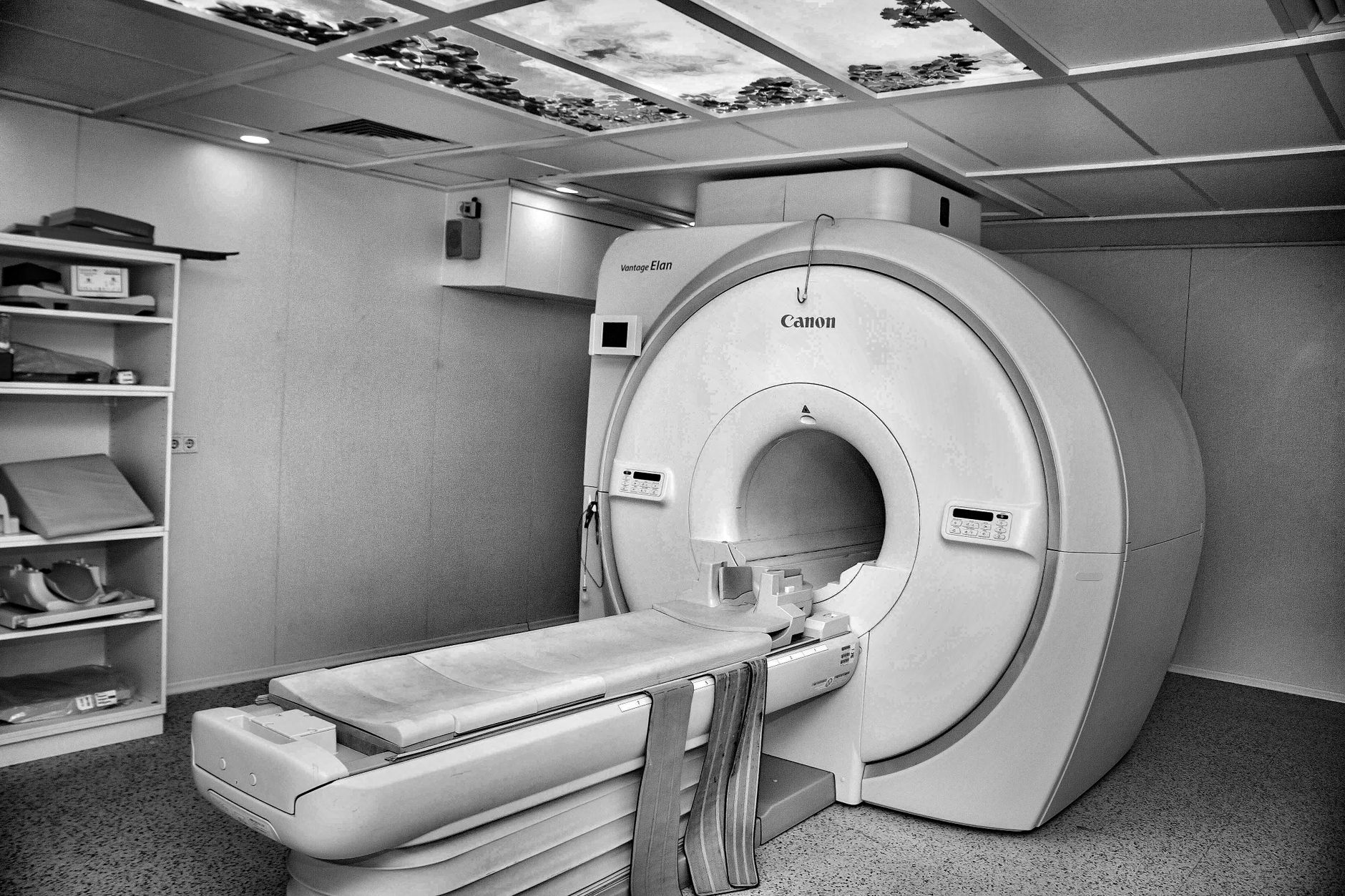
- You will lie on a comfortable, flat table that slides into the MRI machine.
- The machine is a large, open-ended tube — not a fully enclosed box.
- You will hear rhythmic knocking and buzzing sounds. These are completely normal.
- The technologist will ask you to hold your breath for short intervals (usually 10–15 seconds). This keeps the images sharp because the heart and lungs move with breathing.
- Some cardiac MRI scans use a contrast agent (a dye called gadolinium) injected through a small IV in your arm. This highlights certain tissues and is generally very well tolerated.
- The full scan typically takes 45 to 75 minutes.
After the Scan
- If contrast dye was used, you may be asked to drink extra water to help flush it out.
- You can usually go home the same day and resume normal activities.
- A specialist radiologist — often working alongside your cardiologist — will analyse the images and prepare a detailed report.

Is a Cardiac MRI Safe?
For most people, yes — a cardiac MRI is considered very safe. Because it uses magnetic fields and radio waves rather than ionising radiation, it avoids the radiation exposure associated with CT scans or X-rays. This makes it a preferred option when repeated imaging is needed over time, such as monitoring a long-term heart condition.
The gadolinium contrast agent used in some scans is generally safe for people with normal kidney function. If you have kidney problems, your doctor will evaluate this carefully before recommending contrast.
People with certain metal implants — some older pacemakers, cochlear implants, or certain aneurysm clips — may not be able to have an MRI. Always share your complete medical history with your care team before the scan.
Key Takeaways
- A cardiac MRI scan of the heart is a non-invasive, radiation-free test that creates highly detailed images of your heart's structure, function, and tissue health.
- It is recommended when your cardiologist needs information that other tests — ECG, echo, or CT — cannot fully provide.
- Common reasons include assessing heart muscle damage, cardiomyopathy, heart inflammation, valve disease, and congenital conditions.
- The scan takes about 45–75 minutes. You will need to hold your breath briefly at intervals. It is generally comfortable.
- It is considered safe for most people. Metal implants and kidney function should be discussed with your doctor beforehand.
- The results help your cardiologist make precise, personalised treatment decisions — which is exactly why it is recommended.
What Happens After Your Cardiac MRI Results?
Once your cardiologist receives the report, they will discuss the findings with you in detail. The results might confirm a diagnosis, rule out a suspected condition, or guide the next step — whether that is medication, a procedure, or simply closer monitoring.
At the cardiology practice in Rajkot, a cardiac MRI report is always reviewed in the context of your full clinical picture: your symptoms, your history, your other test results, and your personal health goals. A single number or finding is never looked at in isolation.

It is completely natural to feel a little anxious when your doctor recommends a new test. Remember — the reason your cardiologist is asking for this scan is to take better care of your heart, with the most accurate information possible. Knowledge is always on your side.
If you have been advised to undergo a cardiac MRI scan or have questions about any heart test, consider booking a consultation with a qualified interventional cardiologist who can review your individual situation and guide you through the right steps.
Frequently asked questions
- Is a cardiac MRI scan painful or uncomfortable?
- A cardiac MRI is non-invasive and generally comfortable. You will lie still on a padded table, hear some knocking sounds from the machine, and be asked to hold your breath briefly at intervals. If a contrast dye is used, you may feel a mild coolness when it is injected through the IV. Most people find the experience straightforward.
- How long does a cardiac MRI scan take?
- A cardiac MRI typically takes between 45 and 75 minutes from start to finish. The actual imaging time varies depending on how many views and sequences your cardiologist has requested. You can usually go home the same day.
- Can I have a cardiac MRI if I have a pacemaker?
- Many modern pacemakers are MRI-compatible, but this must be confirmed with your cardiologist and the MRI team before the scan is booked. Older pacemaker models and some other metal implants may be a contraindication. Always disclose all implants and past surgeries to your care team.
- How is a cardiac MRI different from an echocardiogram (echo)?
- An echocardiogram uses ultrasound waves and is excellent for a quick, real-time look at heart structure and valve function. A cardiac MRI goes further — it provides more precise measurements, better tissue characterisation (such as detecting scar tissue or inflammation), and superior image quality regardless of body type. Doctors often use both tests together for a complete picture.