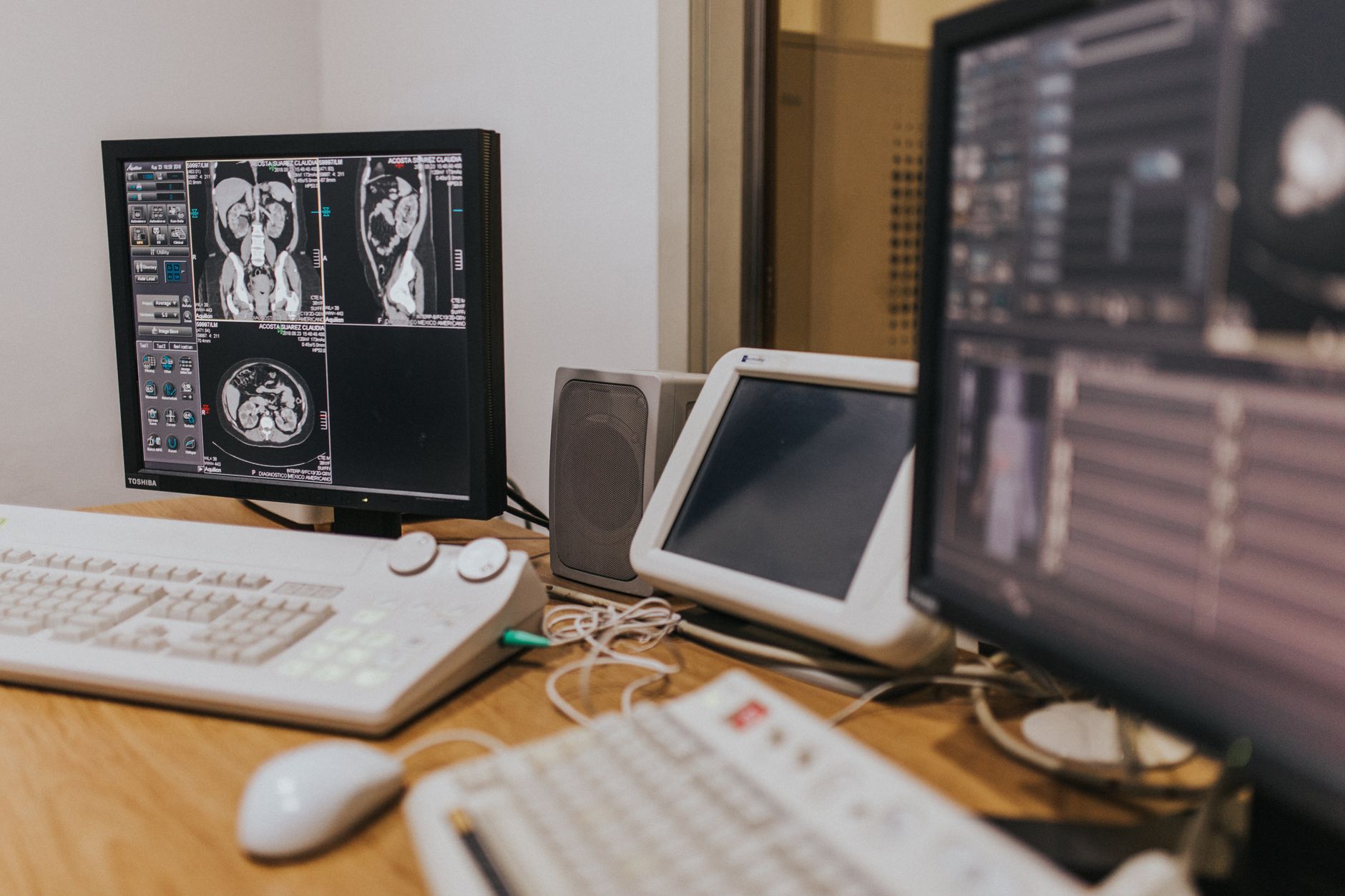
If your cardiologist has suggested a cardiac PET scan and you are wondering what it is, whether it is really necessary, and how it is different from the other heart tests you may have heard of — you are not alone. Many patients in Rajkot and across Gujarat come with exactly these questions. This article explains cardiac PET scan uses in India in plain language, so you can feel informed and confident before your appointment.
What Is a Cardiac PET Scan?
PET stands for Positron Emission Tomography. A cardiac PET scan is a type of nuclear imaging test that takes detailed pictures of how blood flows through your heart muscle and how well that muscle is working at a cellular level.
During the test, a very small, safe amount of a radioactive tracer is injected into a vein in your arm. This tracer travels through your bloodstream and is absorbed by your heart tissue. The PET scanner then detects the energy released by the tracer and creates precise, three-dimensional images of your heart.
The key word here is function. Unlike an X-ray or CT scan that mainly shows the structure of your heart, a cardiac PET scan shows how your heart performs — whether certain areas are receiving enough blood, and whether cells in those areas are still alive and healthy.
How Is It Different from Other Heart Imaging Tests?
There are several imaging tools a cardiologist may use. Understanding how they differ helps explain why a PET scan is sometimes the right choice.
ECG (Electrocardiogram)
An ECG records the electrical activity of your heart. It is quick and non-invasive, but it only captures a snapshot in time and cannot show blood flow in detail.
Echocardiogram (Echo)
An echo uses ultrasound waves to show the heart's structure, size, and movement. It is very useful, but it may miss subtle problems in blood supply to specific regions of the heart muscle.
Treadmill Stress Test (TMT)
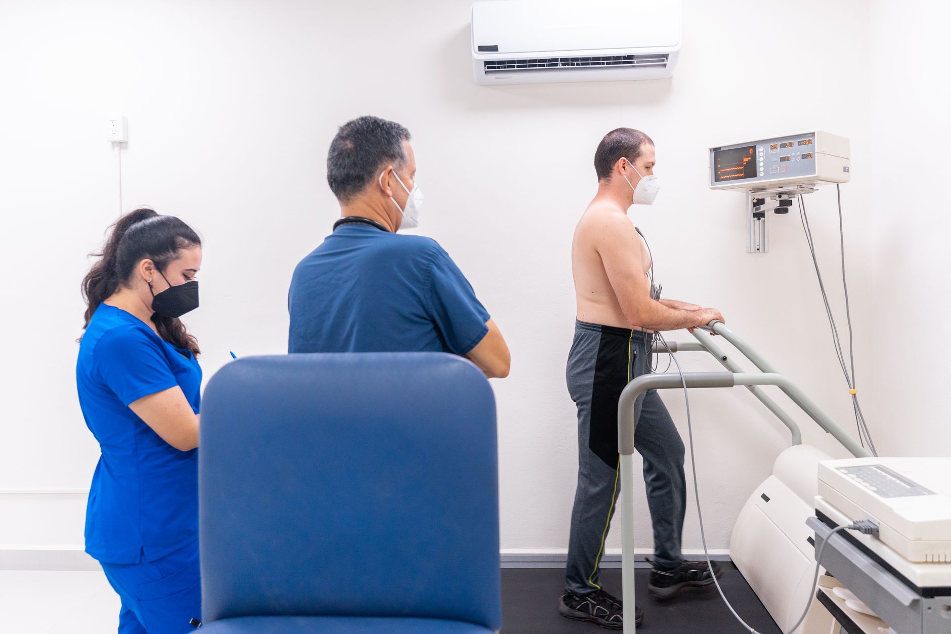
A stress test checks how your heart responds to physical effort. It is a good first-line screen, but it has limitations in certain patients — for example, those who cannot walk on a treadmill due to arthritis or breathing problems, or those whose baseline ECG is abnormal.
SPECT Myocardial Perfusion Imaging (MPI)
SPECT (Single Photon Emission CT) is an older nuclear imaging test that also looks at blood flow in the heart. It has been widely available in India for many years. However, a cardiac PET scan offers higher image quality, shorter scan time, lower radiation exposure, and more accurate measurements of blood flow — especially in patients who are overweight, as body tissue can sometimes blur SPECT images.
CT Coronary Angiography (CTCA)
A CTCA shows the anatomy of the coronary arteries — meaning it can reveal whether there is a narrowing or blockage. But it does not directly tell us whether that narrowing is actually reducing blood flow enough to cause symptoms. A cardiac PET scan answers that follow-up question.
Invasive Coronary Angiography
This is the "gold-standard" test for seeing blockages, but it involves threading a thin tube into the arteries and carries a small procedural risk. A cardiac PET scan can sometimes help doctors decide whether this more invasive step is truly needed.
When Does a Cardiologist Order a Cardiac PET Scan?
A cardiologist will consider a PET scan in specific clinical situations. Here are the most common ones:
1. Diagnosing Coronary Artery Disease (CAD)
When a patient has chest pain or breathlessness and the simpler tests are inconclusive, a cardiac PET scan can accurately identify whether reduced blood flow to the heart muscle (called ischaemia) is the cause. This is one of the primary cardiac PET scan uses in India today.
2. When a Stress Test Result Is Unclear
A 55-year-old patient might have an abnormal stress test result, but the cardiologist needs to know how significant the problem is before recommending further treatment. A cardiac PET scan can precisely map which areas of the heart are affected and how severely.
3. Assessing Heart Muscle Viability Before a Procedure
If a patient has had a heart attack and is being considered for bypass surgery (CABG) or angioplasty, it is important to know whether the damaged heart muscle is still viable — meaning, will it recover if blood supply is restored? This is called myocardial viability assessment, and cardiac PET is regarded as one of the most reliable tools for this purpose.
4. Patients Who Cannot Exercise
For patients with joint pain, severe breathlessness, or other conditions that prevent them from walking on a treadmill, a pharmacological stress PET can be done. A safe medication is used to temporarily increase blood flow to the heart, mimicking the effect of exercise.
5. Obese Patients or Those with a Large Body Frame
As mentioned, body tissue can reduce the accuracy of SPECT scans in heavier patients. The physics of PET imaging are less affected by body size, making it a more reliable option in this group.
6. Monitoring Known Heart Disease
Patients who have already been diagnosed with coronary artery disease and are on medication may need periodic reassessment. A cardiac PET scan can track whether blood flow has improved, stayed the same, or worsened over time.
7. Suspected Cardiac Sarcoidosis or Inflammation
Rare conditions like cardiac sarcoidosis (inflammation of the heart muscle) can be detected with a specific type of cardiac PET scan using a tracer called FDG. This is a specialised use that requires careful patient preparation.
What to Expect on the Day of the Scan
- You will usually be asked to fast for several hours before the test.
- Caffeine (tea, coffee, energy drinks) must be avoided for 24–48 hours beforehand, as it interferes with the blood-flow response.
- The radioactive tracer is injected through a small needle in your arm — the process is straightforward and comfortable.
- You will rest quietly (or undergo pharmacological stress) while the scanner takes images. The entire process typically takes 1–2 hours.
- The radioactivity leaves your body naturally within a few hours, and you can usually return to normal activities the same day.
- Drink plenty of water after the test to help flush the tracer out through urine.

Is a Cardiac PET Scan Safe?
Yes, for most patients it is considered safe. The radiation dose from a modern cardiac PET scan is low — often comparable to or lower than a standard SPECT scan — and the tracers used have a very short half-life, meaning they break down quickly in the body. Your cardiologist will always weigh the benefits of the information gained against any small risks before ordering the test.
Patients who are pregnant are generally not advised to have PET scans unless absolutely necessary. Let your doctor know about all medications and supplements you take before the test.
Cardiac PET Scan Availability in India
Cardiac PET scanning has become increasingly available at advanced cardiac and diagnostic centres in major cities and larger towns across India, including in Gujarat. While it was once only accessible in metro hospitals, growing infrastructure means more patients can now benefit from this technology closer to home.
If you are a patient in Rajkot or the Saurashtra region and your cardiologist has recommended this test, ask specifically about the nearest accredited centre and what preparation you will need.
Key Takeaways
- A cardiac PET scan measures blood flow and heart muscle function — not just anatomy.
- It is particularly useful when simpler tests give unclear results, when assessing heart muscle viability, or when patients cannot exercise.
- PET imaging generally offers higher accuracy than older nuclear tests (SPECT), especially in certain patient groups.
- The test is safe, relatively comfortable, and involves a very short-lived radioactive tracer.
- It helps cardiologists make better-informed decisions about whether a patient needs angioplasty, bypass surgery, or can be managed with medication alone.
- Availability of cardiac PET scanning is growing steadily across India, including in Gujarat.
Every heart is unique, and the right imaging test depends on your individual history, symptoms, and overall health. If you have questions about whether a cardiac PET scan is appropriate for you, please consult a qualified cardiologist. You are welcome to book a consultation with Dr. Nikhila Pachani, Consultant Interventional Cardiologist in Rajkot, to discuss your heart health in detail.
Frequently asked questions
- What is a cardiac PET scan used for?
- A cardiac PET scan is used to measure blood flow to the heart muscle and assess how well the heart tissue is functioning. Common uses include diagnosing coronary artery disease, evaluating heart muscle viability before bypass surgery or angioplasty, and identifying the cause of chest pain when other tests are inconclusive.
- How is a cardiac PET scan different from a stress test or echo?
- A stress test measures how your heart responds to exertion, and an echocardiogram shows the heart's structure and movement. A cardiac PET scan goes further by precisely mapping blood flow through different regions of the heart muscle at a cellular level, providing information that the other two tests cannot.
- Is a cardiac PET scan safe, and how long does it take?
- Yes, a cardiac PET scan is considered safe for most patients. It uses a very small amount of a short-lived radioactive tracer that leaves your body within a few hours. The radiation dose is generally low — often less than or equal to older nuclear heart tests. The full procedure, including rest and stress imaging, typically takes one to two hours.
- Is cardiac PET scanning available in Gujarat and Rajkot?
- Cardiac PET scanning is becoming increasingly available at advanced cardiac and diagnostic centres in Gujarat and across India. If you are in the Rajkot or Saurashtra region, your cardiologist can guide you to the nearest accredited centre and advise you on preparation steps specific to your case.