
You walk into the clinic with a nagging chest discomfort, some breathlessness, or maybe mild ankle swelling. Your doctor listens carefully — and then asks for a chest X-ray. You might wonder: "Isn't an X-ray for the lungs? Why would a cardiologist need one?"
The answer is that a simple, quick, and widely available chest X-ray gives a cardiologist a remarkable amount of information about your heart, the large blood vessels, and the fluid around the lungs — all at once. Think of it as a first window into your chest that helps your doctor decide the next steps.
This article explains, in plain language, exactly what a cardiologist looks for on your chest X-ray — far beyond just checking whether your lungs are clear.
What Is a Chest X-Ray, and How Does It Work?
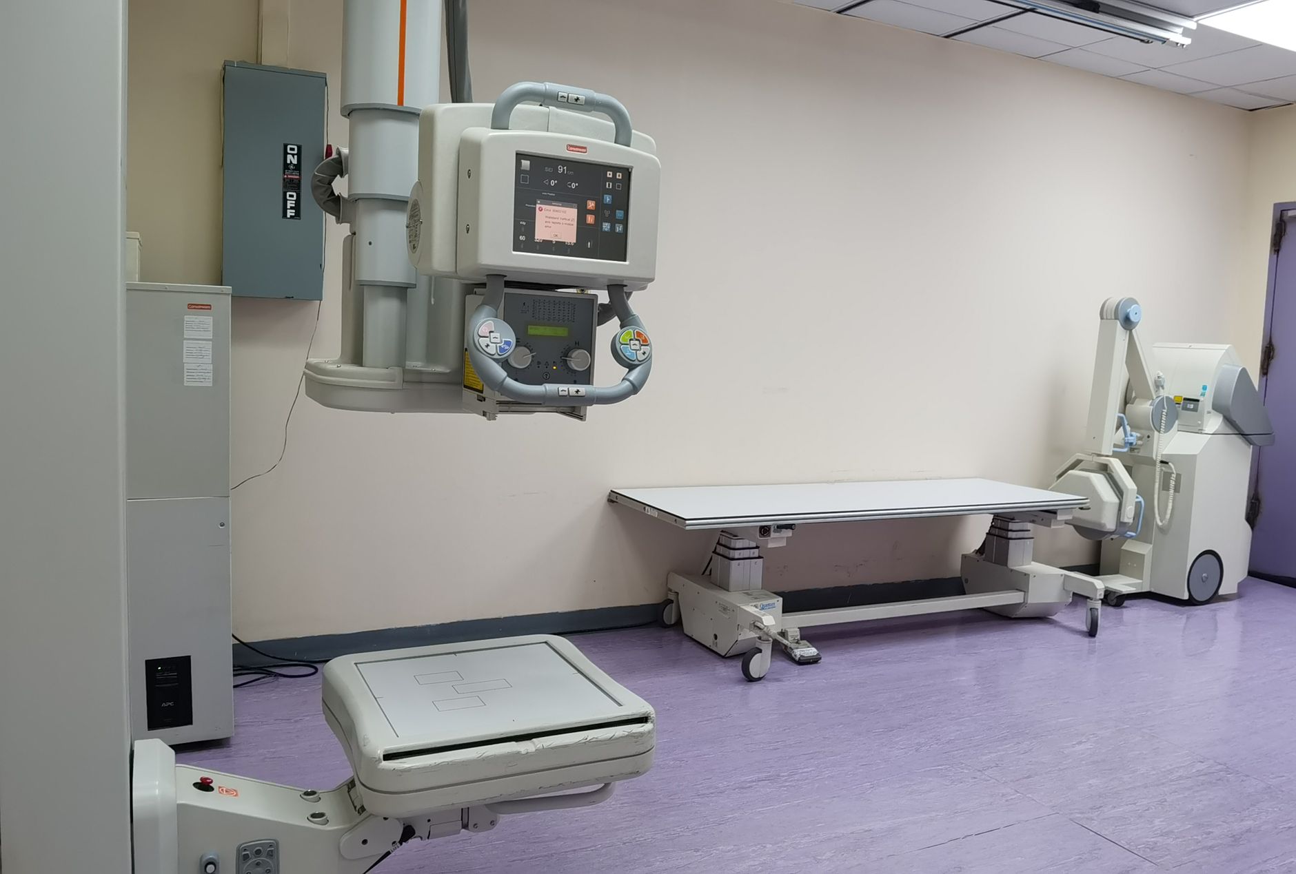
A chest X-ray (also called a CXR) uses a small, carefully controlled beam of radiation to create a shadow-image of everything inside your chest. Dense structures like bones and the heart appear white or light grey. Air-filled lungs appear dark. The result is a flat image that, to a trained eye, tells a rich story.
The whole process takes less than five minutes and is non-invasive and comfortable. You simply stand in front of the X-ray plate, take a deep breath, and hold it for a second. That's it.
In a busy cardiology practice — such as in Rajkot, where many patients come in with undiagnosed breathlessness or chest pain — the chest X-ray is often the very first investigation ordered, because it gives so much information so quickly.
The "Cardiac Silhouette": Reading the Heart's Shadow
On a chest X-ray, the heart casts a shadow called the cardiac silhouette. Cardiologists study this shadow carefully. Here is what they look for:
1. Heart Size (Cardiomegaly)
One of the most important things a cardiologist checks is whether the heart looks enlarged. A standard measurement used is the cardiothoracic ratio (CTR) — the width of the heart compared to the width of the chest. On a standard chest X-ray taken with a full breath in:
- A CTR of less than 0.5 (heart taking up less than half the chest width) is generally considered normal.
- A CTR greater than 0.5 may suggest an enlarged heart, a condition called cardiomegaly.
An enlarged heart does not automatically mean something is dangerously wrong, but it is a flag that prompts further investigation — usually an echocardiogram (heart ultrasound). Cardiomegaly can be seen in conditions like heart failure, a leaky heart valve, or a weakened heart muscle (cardiomyopathy).
2. The Shape of the Heart
The outline of the cardiac silhouette also gives clues. Normally, the heart has a smooth, well-defined border on both sides.
- A boot-shaped heart (coeur en sabot) is a classic X-ray finding in a condition called Tetralogy of Fallot (a congenital heart problem).
- A globular, flask-shaped heart can suggest fluid around the heart — a condition called pericardial effusion.
- Bulging of specific parts of the heart border may indicate problems with individual heart chambers.
For example, a bulge along the left border of the heart might indicate an enlarged left ventricle — the main pumping chamber — which can happen with longstanding high blood pressure or a damaged heart valve.
3. The Aorta and Large Vessels
Just above the heart, the large vessels — particularly the aorta — are visible on the X-ray. Your cardiologist looks at:
- Widening of the aorta, which can suggest aortic dilation or, in urgent situations, an aortic aneurysm.
- Calcification (calcium deposits appearing as bright white lines) in the walls of the aorta, which indicates long-term wear and tear or atherosclerosis.
- The pulmonary arteries (vessels going to the lungs) — if these appear enlarged, it can point to high blood pressure in the lung arteries, called pulmonary hypertension.
Beyond the Heart: What the Lungs Tell the Cardiologist
Here is where many patients are surprised — the lungs on a chest X-ray are actually one of the most important indicators of heart health.
Pulmonary Congestion and Oedema
When the heart is not pumping efficiently, blood can back up into the blood vessels of the lungs. This is called pulmonary congestion. On an X-ray, this appears as:
- Prominent (engorged) blood vessels in the upper parts of the lungs — a sign called "upper lobe diversion" or "upper lobe venous engorgement."
- Haziness or a "bat-wing" pattern spreading from the centre of the chest outward — a sign of pulmonary oedema (fluid leaking into the lung tissue itself).
- Kerley B lines — tiny horizontal white lines at the outer edges of the lungs, caused by fluid in the small channels between lung segments. These are a classic sign of heart failure.
A 55-year-old with a history of high blood pressure who comes in with worsening breathlessness at night might have an X-ray that shows all of these features — upper lobe engorgement, haziness, and Kerley B lines — pointing clearly toward heart failure even before any blood test result is available.
Pleural Effusion
Pleural effusion means fluid has accumulated in the space between the lung and the chest wall. While it can happen for many reasons (infections, kidney disease), in a cardiology setting it is often caused by heart failure. On an X-ray, it appears as a blunting (filling in) of the normally sharp angle at the base of the lung, or as a white haze at the bottom of one or both lungs.
Bilateral pleural effusions (fluid on both sides) are a strong clue toward a cardiac cause.
Other Important Cardiac Clues on a Chest X-Ray
Prosthetic Valves and Pacemakers
If a patient has had heart surgery or a procedure before, the chest X-ray can confirm:
- The position and type of prosthetic (artificial) heart valves — these appear as distinct metallic rings or structures.
- The position of pacemaker leads and the pacemaker device itself — ensuring the leads are correctly placed in the right chambers of the heart.
- Surgical clips or wires from previous bypass surgery.
This is particularly useful when a patient moves to a new city — as many people do when coming to Rajkot from smaller towns across Saurashtra — and their old records are not immediately available.
Pulmonary Hypertension Signs
Apart from enlarged pulmonary arteries, the X-ray may show a prominent main pulmonary artery segment (a visible bulge just to the left of the upper heart border). Combined with enlarged right heart chambers, this pattern raises concern for pulmonary hypertension and warrants further testing.

What a Chest X-Ray Cannot Tell You
It is equally important to understand the limits of a chest X-ray. It is a snapshot — helpful but incomplete. It cannot:
- Show the internal structure or function of the heart valves.
- Detect blockages in the coronary arteries (for that, you need tests like a stress test, CT coronary angiography, or invasive coronary angiography).
- Measure the pumping strength (ejection fraction) of the heart — an echocardiogram is needed for that.
- Pick up early or subtle changes that haven't yet affected heart size or lung appearance.
So while the chest X-ray is an excellent starting point, it is almost always followed by further investigations based on what it shows — or doesn't show.
Key Takeaways
- ✅ A chest X-ray is a quick, non-invasive first step that reveals important heart clues, not just lung findings.
- ✅ Cardiologists study the cardiac silhouette for heart size, shape, and the appearance of large vessels like the aorta.
- ✅ Lung changes on an X-ray — like pulmonary congestion, Kerley B lines, and pleural effusion — are strong indicators of how well the heart is working.
- ✅ The X-ray can also confirm the position of pacemakers, leads, and prosthetic valves.
- ✅ A chest X-ray is a starting point, not an endpoint — your cardiologist will use its findings to guide further, more detailed investigations.
- ✅ Never ignore an abnormal chest X-ray report. Discuss every finding with your cardiologist for proper context.
Frequently Asked Questions
See the FAQ section below for answers to common questions about chest X-rays and heart health.
If you have received a chest X-ray report with unfamiliar findings, or if you have been experiencing chest discomfort, breathlessness, or palpitations, we encourage you to speak with a qualified cardiologist who can review your reports in the context of your full health picture. You are welcome to book a consultation with Dr. Nikhila Pachani at her Rajkot clinic for a thorough, personalised cardiac assessment.
Frequently asked questions
- Can a chest X-ray detect a heart attack?
- Not directly. A chest X-ray cannot show blocked coronary arteries or detect a heart attack in progress. However, it can reveal complications that may follow a heart attack, such as an enlarged heart or fluid buildup in the lungs. An ECG and blood tests (cardiac enzymes) are the primary tools for diagnosing a heart attack.
- Is the radiation from a chest X-ray safe?
- Yes, the radiation dose from a single chest X-ray is very low — roughly equivalent to the natural background radiation you receive over a few days of normal life. It is considered safe for most adults. Your doctor will always weigh the benefit of the information gained against any minimal risk.
- If my chest X-ray looks normal, does that mean my heart is healthy?
- Not necessarily. A normal chest X-ray is reassuring, but it does not rule out all heart conditions. Coronary artery disease, early valve problems, and abnormal heart rhythms may not show any changes on a chest X-ray. Your cardiologist may still recommend further tests like an ECG or echocardiogram based on your symptoms.
- How is a chest X-ray different from an echocardiogram?
- A chest X-ray produces a still shadow-image that shows the overall size and shape of the heart and changes in the lungs. An echocardiogram uses sound waves (ultrasound) to create a moving, detailed image of the heart's internal chambers, valves, and pumping function. They provide different and complementary information — your cardiologist often uses both together.