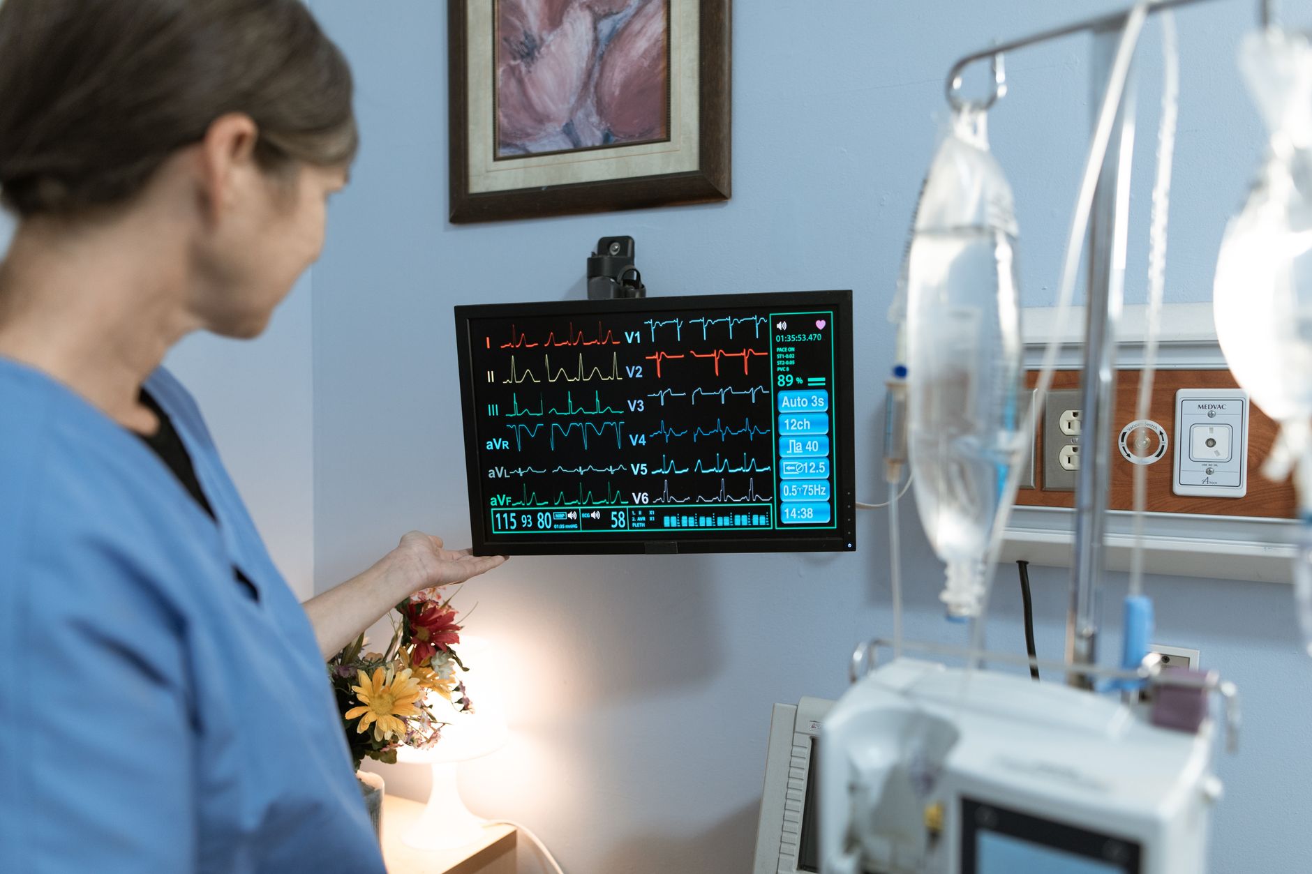
What Is Fluid Around the Heart?
Your heart is not simply floating freely inside your chest. It sits inside a thin, double-layered protective sac called the pericardium. Under normal, healthy conditions, there is a very small amount of fluid — typically between 15 and 50 millilitres — between the two layers of this sac. This tiny cushion of fluid helps reduce friction as the heart beats steadily, day after day.
Sometimes, however, this fluid builds up in larger amounts than normal. When that happens, the condition is called a pericardial effusion. In many cases, a mild pericardial effusion causes no major problems and can be monitored carefully over time.
But when the fluid accumulates rapidly or builds up to a significant volume, it starts to squeeze the heart from the outside — preventing the heart's chambers from filling properly with blood. This serious, life-threatening condition is known as fluid around the heart cardiac tamponade, or simply cardiac tamponade.
Understanding what tamponade is, why it happens, and how it is treated can help you or your loved ones recognise warning signs and seek timely care.
Why Does Fluid Build Up Around the Heart?
There are several reasons why excess fluid may collect inside the pericardial sac. Some of the most common causes include:
- Infections — Viral infections (such as those that cause pericarditis, an inflammation of the heart sac) are among the most frequent culprits, especially in younger, otherwise healthy individuals.
- Cancer — Certain cancers, including lung cancer, breast cancer, and lymphoma, can spread to the pericardium and trigger fluid accumulation.
- Autoimmune conditions — Diseases such as lupus or rheumatoid arthritis can cause inflammation of the pericardium.
- Kidney failure — Advanced kidney disease can lead to a build-up of waste products that irritate the pericardium.
- Heart attack or heart surgery — In some cases, bleeding into the pericardial sac can follow a heart attack, an aortic dissection, or a cardiac procedure.
- Tuberculosis (TB) — TB-related pericardial effusion is particularly relevant in India, including here in Gujarat, where TB remains an important public health concern.
- Hypothyroidism — An underactive thyroid gland is another recognised cause.
- Trauma — A significant injury to the chest can result in bleeding around the heart.
In some patients, no clear cause is found — this is called an idiopathic effusion.
How Cardiac Tamponade Develops
Think of the pericardium as a firm, relatively non-elastic bag. When fluid accumulates slowly over weeks or months, the pericardium gradually stretches to accommodate it — sometimes up to a litre or more — before symptoms appear. This is why some large effusions are discovered incidentally during a routine scan and cause only mild discomfort.
However, when fluid accumulates quickly — even a relatively small amount, say 150–200 ml — the pericardium has no time to stretch. The rising pressure inside the sac rapidly compresses the heart's chambers. The right side of the heart, which is under lower pressure, is squeezed first. Blood cannot fill the heart properly. The heart's pumping output drops sharply. Blood pressure falls. The body's vital organs begin to receive less blood and oxygen.
This is cardiac tamponade — a medical emergency that requires prompt recognition and treatment.
Recognising the Warning Signs
Symptoms of fluid around the heart and cardiac tamponade can vary depending on how quickly the fluid has built up. The classic warning signs doctors look for include:
- Breathlessness — feeling short of breath even at rest, or when lying flat
- Chest discomfort or pressure — a heavy, tight, or aching feeling in the chest
- Rapid heartbeat (palpitations) — the heart tries to compensate by beating faster
- Dizziness or light-headedness — due to low blood pressure and reduced blood flow to the brain Fatigue and weakness — a feeling of exhaustion that comes on relatively quickly
- Swelling of the neck veins — a visible sign that blood is backing up because the heart cannot pump effectively
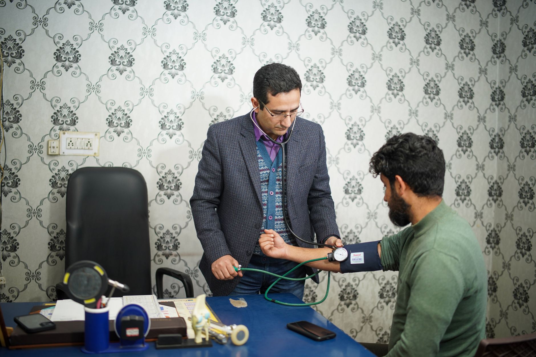
In a medical emergency setting, doctors are trained to look for the "Beck's Triad" — three classic findings: low blood pressure, distended (swollen) neck veins, and muffled or distant heart sounds on a stethoscope.
Important: If you or someone around you experiences sudden severe breathlessness, chest pain, dizziness, and a feeling of faintness, seek emergency medical care immediately. Do not wait.
How Is Cardiac Tamponade Diagnosed?
Diagnosing tamponade requires both clinical assessment and a few targeted investigations.
Echocardiogram (Echo)
This is the most important and reliable diagnostic tool. An echocardiogram uses sound waves to create a real-time image of the heart. It can clearly show:
- The presence and size of the pericardial effusion
- Compression (collapse) of the right-sided heart chambers — a key sign of tamponade
- How severely the heart's pumping function is affected
Chest X-Ray
A chest X-ray may show an enlarged, balloon-shaped heart shadow when a large effusion is present. However, it may appear normal if the fluid has built up rapidly.
Electrocardiogram (ECG)
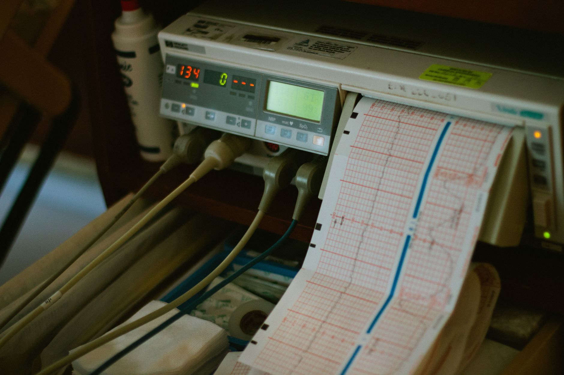
An ECG can reveal characteristic changes — particularly a pattern called electrical alternans, where the height of the heartbeat tracings alternates with every beat. This happens because the heart is swinging back and forth within the fluid-filled sac.
CT Scan or MRI
These imaging tests may be used to better understand the cause of the effusion and its relationship to surrounding structures.
Treatment: Relieving the Pressure
Once cardiac tamponade is confirmed or strongly suspected, relieving the pressure around the heart becomes the immediate priority. There is one primary procedure for this:
Pericardiocentesis
Pericardiocentesis is the procedure of draining the excess fluid from around the heart using a thin needle or catheter, guided by echocardiography or X-ray imaging. In experienced hands, this procedure is performed carefully and effectively, providing rapid relief.
Once the fluid is drained, blood pressure typically improves quickly, and patients usually feel significantly better within a short time. A small drainage catheter may be left in place temporarily to allow any remaining fluid to drain over the following day or two.
The drained fluid is sent for laboratory analysis — this helps identify whether infection, cancer cells, TB, or other causes are responsible, guiding further treatment.
Surgical Drainage
In certain situations — for example, when the effusion recurs repeatedly, or when the pericardium is thickened and scarred (constrictive pericarditis) — a surgeon may create a small opening in the pericardium (called a pericardial window) to allow continuous drainage. This is less commonly required but is an important option for selected patients.
Treating the Underlying Cause
Draining the fluid addresses the immediate emergency, but treating the root cause is equally important. For example:
- Viral pericarditis is usually managed with anti-inflammatory medications (such as NSAIDs and colchicine)
- TB-related effusion requires a full course of anti-tuberculosis therapy
- Cancer-related effusion may need oncology treatment in addition to drainage
- Autoimmune causes are managed with appropriate disease-modifying therapy
Living After Cardiac Tamponade
Most patients who receive prompt, appropriate treatment recover well. The long-term outlook depends largely on the underlying cause. For instance:
- A young person whose tamponade was caused by a viral infection often makes a full recovery with no lasting cardiac damage.
- A 55-year-old patient whose effusion was related to an autoimmune condition, for example, will need ongoing monitoring and treatment for that condition, but can live a full, active life with proper follow-up care.
Regular follow-up echocardiograms are recommended to ensure the fluid does not re-accumulate. Your cardiologist will also advise on activity, medications, and any lifestyle adjustments based on your specific situation.
Key Takeaways
- The heart sits inside a protective sac (the pericardium) that normally contains a small amount of fluid.
- When excess fluid builds up rapidly, it can compress the heart — a life-threatening emergency called cardiac tamponade.
- Common causes in India include viral infections, tuberculosis, cancer, kidney disease, and autoimmune conditions.
- Warning signs include breathlessness, chest discomfort, rapid heartbeat, dizziness, and swollen neck veins.
- An echocardiogram is the most important tool for diagnosis.
- Pericardiocentesis — draining the fluid under image guidance — is the primary treatment and provides rapid relief.
- Identifying and treating the underlying cause is essential for long-term recovery.
- Most patients recover well with timely intervention and appropriate follow-up.
If you are experiencing chest discomfort, unexplained breathlessness, or have been told you have fluid around the heart, please do not delay seeking specialist advice. Consult a qualified cardiologist for a thorough evaluation — early assessment can make a real difference to your outcome.
Frequently asked questions
- What is the difference between a pericardial effusion and cardiac tamponade?
- A pericardial effusion simply means there is extra fluid around the heart inside the pericardial sac. This can range from mild to large, and may not cause major symptoms if it builds up slowly. Cardiac tamponade occurs when the fluid — particularly if it accumulates rapidly — creates enough pressure to compress the heart and seriously impair its ability to pump blood. Tamponade is a medical emergency, while a small, stable effusion may only need careful monitoring.
- How quickly does cardiac tamponade develop?
- It depends on how fast the fluid accumulates. When fluid builds up slowly over weeks or months (for example, in some cancers or autoimmune conditions), the pericardium gradually stretches and tamponade may develop late or not at all. But when fluid accumulates rapidly — such as after a heart injury or bleeding — even a relatively small amount (150–200 ml) can cause tamponade within minutes to hours, making it an urgent emergency.
- Is pericardiocentesis a major operation?
- Pericardiocentesis is a minimally invasive procedure rather than open surgery. A cardiologist inserts a thin needle or catheter through the chest wall to drain the fluid, guided by echocardiography or X-ray imaging to ensure precision and safety. Most patients experience significant relief in breathlessness and discomfort soon after the fluid is drained. It is routinely performed in well-equipped cardiac centres.
- Can cardiac tamponade come back after treatment?
- Yes, in some patients the fluid may re-accumulate, especially if the underlying cause — such as cancer, tuberculosis, or an autoimmune condition — has not been fully treated. This is why follow-up echocardiograms and treatment of the root cause are so important after the initial drainage. In cases of recurrent effusion, a procedure called a pericardial window may be considered to provide a more lasting solution.