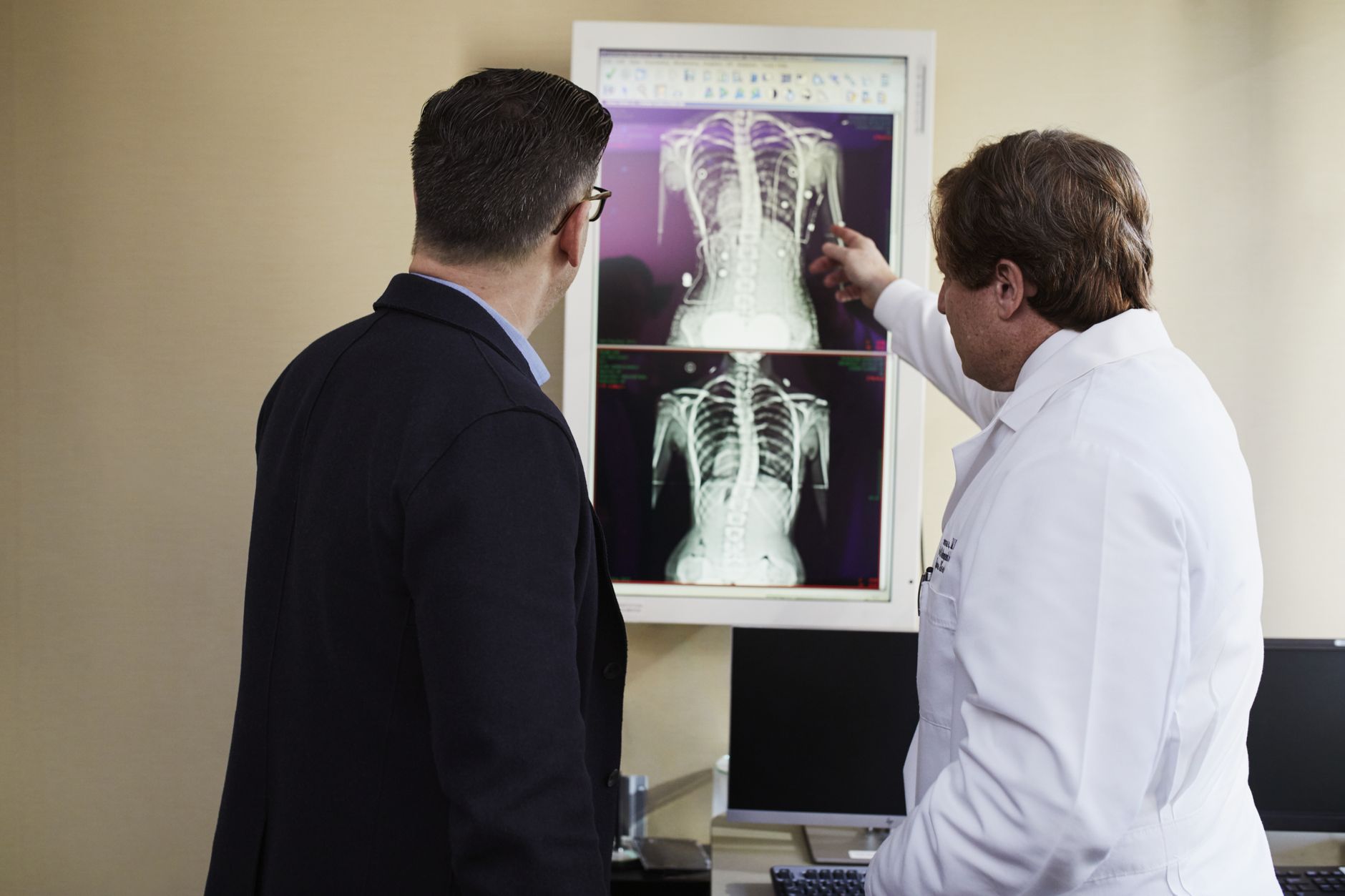
What Is a Patent Foramen Ovale (PFO)?
Before you are born, your heart works differently from how it will after birth. Inside the womb, a baby's lungs are not yet used for breathing — the placenta does that work instead. To allow blood to bypass the lungs, nature creates a small opening between the two upper chambers of the heart (the right and left atria). This opening is called the foramen ovale.
At birth, when a baby takes its first breath and the lungs begin to work, this opening is no longer needed. In most people, it seals shut on its own within the first year or two of life. But in roughly one in four adults — about 25% of the population — the opening never fully closes. When it stays open, it is called a patent foramen ovale, or PFO.
The word "patent" simply means "open." So a PFO is literally a small, flap-like channel that remains between the two sides of the heart. For the vast majority of people, a PFO causes no symptoms whatsoever and is discovered only by chance during a heart scan for another reason.

So What Is the Problem?
A PFO on its own is usually harmless. The flap acts almost like a one-way valve — under normal conditions, blood pressure on the left side of the heart keeps it firmly shut.
The concern arises in specific situations — for example, during a Valsalva manoeuvre (straining, coughing forcefully, or bearing down). At that moment, pressure in the right atrium can briefly spike above the left, causing the flap to pop open momentarily. If a tiny blood clot or air bubble happens to be floating in the right side of the heart at that instant, it can cross over to the left side and travel up to the brain — potentially causing a stroke or transient ischaemic attack (TIA).
Understanding Cryptogenic Stroke
Doctors use the term "cryptogenic stroke" for a stroke whose cause cannot be found despite thorough investigation. No blocked artery, no irregular heart rhythm (like atrial fibrillation), no other obvious explanation. Studies consistently show that PFO is found far more often in people who have had a cryptogenic stroke — especially younger adults — compared with the general population.
This is the heart of the patent foramen ovale stroke connection: a small unnoticed hole may be quietly acting as a trapdoor, allowing clots to sneak from the venous circulation into the arterial circulation and reach the brain.
Who Is Most at Risk?
Not every person with a PFO will ever have a stroke, and not every stroke in a person with a PFO is caused by the PFO. Cardiologists weigh several factors:
- Size of the PFO — larger openings carry a higher risk of clot passage
- Presence of an atrial septal aneurysm (ASA) — a floppy, bulging wall near the PFO that further increases risk
- Amount of right-to-left shunting — how much blood (or bubbles) actually crosses over
- Age and stroke history — a cryptogenic stroke in someone under 60 raises stronger suspicion of PFO
- Deep vein thrombosis (DVT) or clotting tendency — clots from leg veins can travel to the heart
In a city like Rajkot, where lifestyle factors such as high-salt diets, sedentary habits, and uncontrolled diabetes are common, the overall cardiovascular risk picture can make a PFO more clinically significant — which is why a thorough individual evaluation always matters.

How Is PFO Diagnosed?
Several tests help doctors identify a PFO and understand how significant it is:
Bubble Echocardiogram (Bubble Echo)
This is the most common test. A saline solution mixed with tiny air bubbles is injected into a vein. The doctor watches on an ultrasound whether bubbles cross from the right to the left side of the heart. Crossing bubbles strongly suggest a PFO.
Transoesophageal Echocardiogram (TOE/TEE)
A small ultrasound probe is gently passed into the food pipe (oesophagus) to get very detailed images of the heart's walls and chambers. This gives much clearer pictures than a standard chest echo and can precisely measure the PFO.
Transcranial Doppler (TCD)
Using ultrasound on the skull, this test detects bubbles reaching the brain's blood vessels — a sensitive way to confirm a right-to-left shunt.
Brain MRI
After a suspected stroke or TIA, a brain MRI helps confirm whether any damage has occurred and guides decisions about treatment urgency.
What Are the Treatment Options?
Treatment decisions are highly individualised. An illustrative example: a 42-year-old who has had a cryptogenic stroke and is found to have a large PFO with an atrial septal aneurysm would likely be considered for closure, whereas an otherwise healthy 35-year-old with a tiny incidental PFO and no symptoms might simply be monitored.
1. Medications (Blood Thinners or Antiplatelets)
For many patients — especially those with small PFOs, older age, or additional risk factors — antiplatelet medicines (such as aspirin) or anticoagulants (blood-thinning medicines) are prescribed to reduce the risk of clot formation. This approach avoids any procedure but requires long-term medication compliance.
2. Percutaneous PFO Closure (Catheter-Based Procedure)
For carefully selected patients — particularly those with a significant PFO and a history of cryptogenic stroke — transcatheter PFO closure is a well-established, minimally invasive option. Here is how it works:
- A thin, flexible tube (catheter) is guided through a vein in the groin up to the heart
- A small, umbrella-like closure device is placed across the PFO
- Over weeks to months, the heart's own tissue grows over the device, permanently sealing the hole
- Most patients go home the same day or the next day
Large randomised clinical trials (RESPECT, CLOSE, GORE-REDUCE, and others) have shown that PFO closure significantly reduces the risk of recurrent stroke in properly selected younger patients with cryptogenic stroke compared with medication alone.
3. Ongoing Monitoring
For asymptomatic individuals discovered to have a PFO during routine scans, watchful waiting with periodic follow-up is often entirely appropriate. The vast majority never develop any related problem.
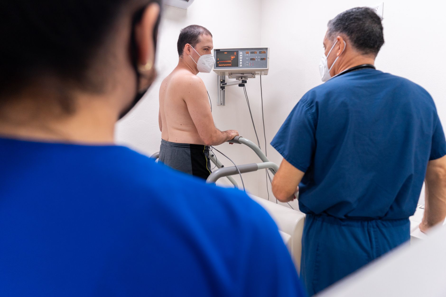
Living With a PFO: What You Can Do
If you have been diagnosed with a PFO — with or without a stroke history — here are sensible everyday steps:
- Stay active — regular, moderate exercise supports heart health; always discuss intensity levels with your doctor first
- Manage underlying risk factors — blood pressure, blood sugar, and cholesterol should be well-controlled
- Stay hydrated — dehydration can thicken the blood and promote clot formation
- Report new symptoms promptly — sudden weakness on one side, slurred speech, vision changes, or a severe headache should never be ignored
- Discuss long-haul flying — prolonged immobility can raise DVT risk; your doctor may advise compression stockings or movement breaks
- Avoid self-medicating — never start or stop blood thinners without medical guidance
Key Takeaways
- A patent foramen ovale (PFO) is a small, persistent opening between the heart's upper chambers, present in about 1 in 4 adults
- Most people with a PFO have no symptoms and live completely normal lives
- The patent foramen ovale stroke connection is strongest in cryptogenic (unexplained) stroke, particularly in adults under 60
- Diagnosis involves a bubble echocardiogram, TOE, or transcranial Doppler, often combined with brain MRI
- Treatment ranges from medications to a minimally invasive catheter-based closure procedure, depending on individual risk
- Not all PFOs need treatment — a thorough cardiologist evaluation is essential before any decision is made
This article is intended for general patient education and does not constitute individual medical advice. If you or a family member has experienced an unexplained stroke, TIA, or has been told about a PFO, consider booking a consultation with a qualified interventional cardiologist for a personalised evaluation and guidance tailored to your specific situation.
Frequently asked questions
- Can a patent foramen ovale cause a stroke without any warning signs?
- Yes, in many cases a PFO-related stroke can occur without any prior symptoms related to the heart defect itself. The PFO may have been present since birth and completely unnoticed. This is why a cryptogenic (unexplained) stroke — especially in a younger adult — often prompts doctors to specifically look for a PFO as a possible underlying cause.
- Is PFO closure a major open-heart surgery?
- No. PFO closure today is typically done as a minimally invasive, catheter-based (transcatheter) procedure. A thin tube is guided through a vein in the groin to the heart, and a small closure device is placed — no open-chest surgery is needed. Most patients are discharged within one to two days and return to normal activities relatively quickly.
- If I have a PFO and have never had a stroke, do I still need treatment?
- Not necessarily. The majority of people with an incidental, asymptomatic PFO do not require any intervention and simply need periodic monitoring. Treatment decisions depend on the size of the PFO, associated features (like an atrial septal aneurysm), personal risk factors, and medical history. Your cardiologist will weigh all these factors to recommend the most suitable approach for you.
- How is a PFO different from a hole in the heart (ASD)?
- Both involve an opening between the heart's upper chambers, but they are different conditions. An atrial septal defect (ASD) is a true structural gap in the heart's wall that is usually present from birth and often larger. A PFO is a flap-like channel — the remnant of a normal fetal structure — that failed to seal after birth. PFOs are generally smaller and behave differently from ASDs, though both can be diagnosed and treated by an interventional cardiologist.